Methods & Techniques
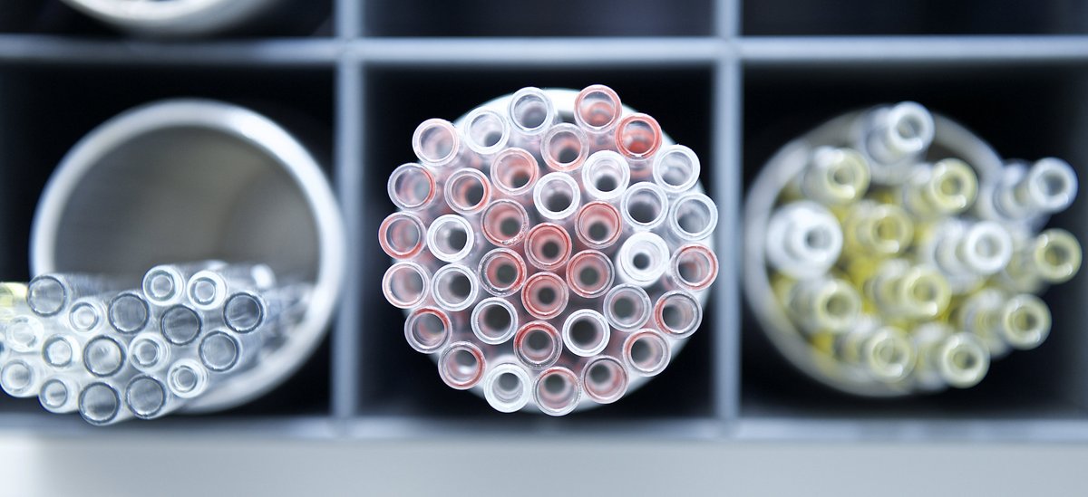
For our research on proteostasis in bacteria, yeast cells and C. elegans worms we apply a broad variety of methods and techniques.
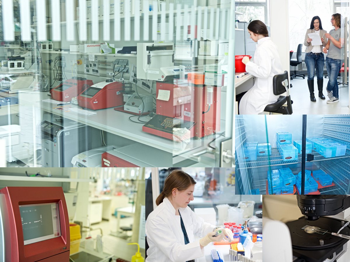
In November 2014 we moved into a brandnew research building where we have all the necessary equipment at hand to do state-of-the-art biochemistry and genetics. Recently, we expanded our spectrum of methods to analyze the interaction of molecules by the microscale thermophoresis technique.
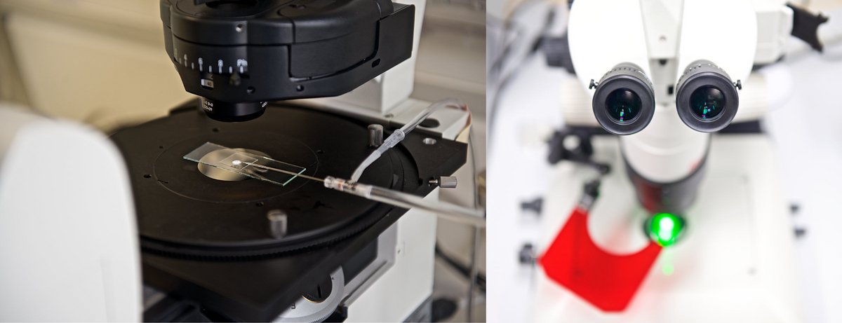
We produce transgenic C. elegans worms either by using CRISPR/Cas9 for gene editing or by injecting plasmids carrying genes of interest to obtain transgenic animals carrying extrachromosomal arrays. For microinjection into the gonads we have two inverted microscope stands equipped with DIC in our lab. These stands have cameras attached to facilitate training of new lab members and of students who wish to learn how to microinject worms. With our fluorescence stereo microscopes we identify transgenic progeny after microinjection of worms and use them to record their movement.
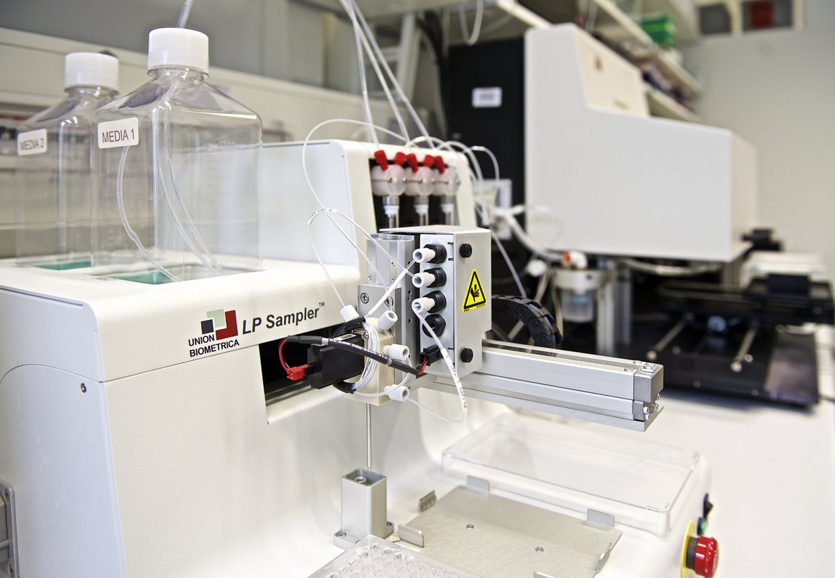
Worms carrying extrachromosomal arrays are separated from non-transgenic animals with the help of our worm sorter, the Copas FlowPilot. This instrument allows to rapidly and very gently sort thousands of animals at different stages of development. Afterwards the sorted animals are either used for biochemical experiments or incubated further.
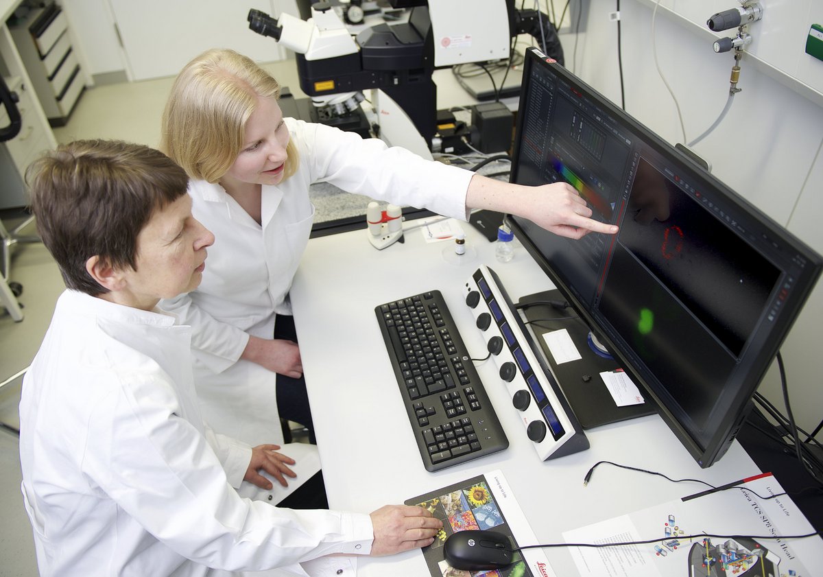
Research in our lab also depends on high-quality imaging of our model organisms. Our confocal microscope SP8 equipped with several sensitive HyD detectors and a HyVolution module allows to measure the subcellular distribution of a particular molecule of interest at superresolution level.
The University of Konstanz´s Department of Biology has several core facilities where we also perfom measurements: We do mass spectrometric analyses at the Department of Biology´s Proteomics Core Facility and high throughput screening at the University´s Screening Centre. Besides standard confocal imaging the BioImaging Center also allows to do superresolution 3D SIM imaging. Suspensions of cells can be sorted at the Flow Cytometry Center and the Electron Microscopy Center of the Department of Biology offers the opportunity to analyze samples with various electron microscopic techniques.