Zeiss LSM 880 with AiryScan
The Zeiss LSM880 is a point laser scanning confocal microscope equipped with a highly sensitive GaAsP detector for spectral imaging and the novel AiryScan detector with a high signal to noise ratio which allows to image at 0.2 Airy unit resolution. The fast mode of the AiryScan detector greatly improves imaging speed, and is especially suited to image large 3D volumes or live samples.
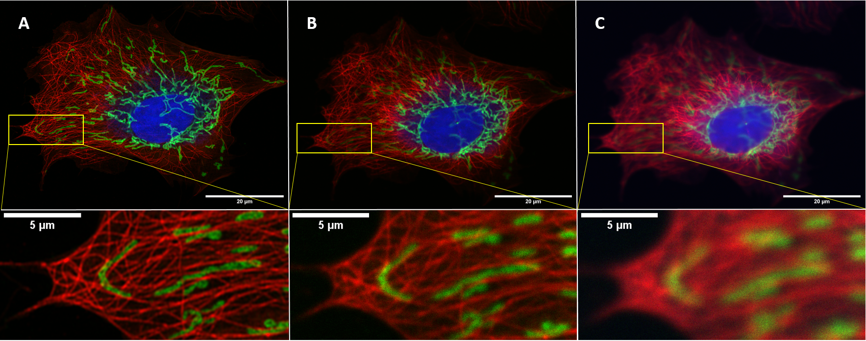
This image comparison showcases the differences between airy-scan (A), confocal (B), and non-confocal (C) microscopy techniques. Using fluorescent dyes, different structures of a cell are labeled, with the nucleus stained in blue, mitochondria in green, and tubulin in red.
Non-confocal imaging involves setting the pinhole to its maximum size, which allows light from out-of-focus planes to contribute to the image (C). However, by closing the pinhole to a size of about 1 airy unit, out-of-focus light is eliminated, resulting in increased contrast and slightly improved resolution in confocal images (B).
Further closing the pinhole improves the resolution of the image, but at the cost of reduced signal detection. The Airy Unit detector offers a solution to this problem, with 32 GaAsP detector elements arranged in a 1.25 AU compound eye pattern. Each element behaves like a small 0.2 AU pinhole, combining increased resolution with good signal detection. With an additional deconvolution step, the Airyscan provides 1.7x higher resolution in all three spatial dimensions than conventional laser scanning confocal microscopy (A).

Confocal microscopy provides a significant advantage over traditional microscopy by enabling optical sectioning of samples and generating 3D structures without the need for physical dissection. By scanning the sample in the x-y plane at a known depth, a process known as optical sectioning, a series of horizontal sections, or Z-stacks, are obtained. By changing the z-axis focus each time, the depth at which the microscope scans is altered by moving the stage, resulting in a collection of images used to calculate and generate the 3D structure of the sample. This feature is invaluable in understanding and analyzing the complete structure of a sample.