Research Projects
Our research focus is axon regeneration and the role of the proteins Reggie/Flotillin, Thy-1 and Nogo.
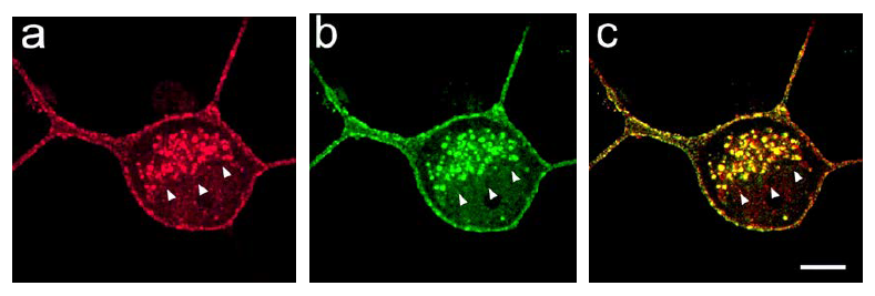
Reggies promote the targeted delivery of cargo molecules to distinct sites of the cell
Reggies are commonly known as microdomain/lipid raft proteins which communicate with the GPI-anchored proteins PrP and Thy-1 during axon growth. All are upregulated in fish retinal ganglion cells during retinal axon regeneration after lesion to the optic nerve. Downregulation of the Reggies blocks regeneration as well as neuronal differentiation in mammalian hippocampal neurons.
Recent work demonstrates that Reggies interact with Rab11 during cargo trafficking and promote the targeted delivery of functionally important membrane proteins to specific sites of the cell. Reggies are involved in the delivery of E-cadherin to adherens junctions, integrins to focal adhesions, the T cell receptor to the T cell cap, growth-related proteins to the growth cone of elongating axons.
Recently, we found that Reggies participate in the targeted recycling of PSD-95 and glutamate receptors at the postsynaptic site of spines in hippocampal neurons. Our results suggest that Reggies promote synaptic plasticity by regulating together with Rab11 the delivery of postsynaptic cargo to spines (Hülsbusch at al., 2015; Solis et al., 2013; Bodrikov et al., 2017), For further information
Nogo/Rtn4b is expressed at low levels in the lesioned zebrafish optic nerve but is upregulated in RGCs to support axon regeneration
Rtn4a and Rtn4b have recently been identified as the zebrafish homologues of mammalian Nogo-A. Nogo-A is a potent inhibitor of axon regeneration in the CNS of mammals. Zebrafish Rtn4b has a Nogo-A specific region which can block axon growth. However, retinal ganglion cell axons regenerate successfully in zebrafish apparently because of very low level expression of Rtn4b in the myelin of the optic nerve. A quite different function was observed for Rtn4b as an ER-associated protein within neurons. Rtn4b is massively upregulated in the retinal ganglion cells after optic nerve lesion and promotes axon regeneration (as an ER associated protein) (Bodrikov et al., 2017; Welte at al., 2015).
Thy-1 is upregulated by retinal ganglion cells and supports axon regeneration
Thy-1 is upregulated in goldfish and zebrafish retinal ganglion cells after optic nerve lesion. We have recently discovered that Thy-1 promotes axon regeneration. These findings together with new insights into the role of Thy-1 in mammalian neurons and the participation of Reggies in Thy-1 induced signaling (unpublished) identify Thy-1 as a positive effector of neurite elongation.